Often, a foot pain diagnosis can help in bringing up an articular cartilage injury, as articular cartilage damage is not similar to other body part damages. There is a chance that the affected people will never realize that they have damaged their cartilage, as it does not show immediately in any physical form.
Foot Pain Diagnosis Knowhow

There are no specific tests that are used to determine the extent of damage that the articular cartilage has gone through. Therefore, unlike ligament tears, cartilage repair takes a considerable amount of effort both on the part of the doctor who’s administering the treatment and the patient. There are a few things that will help a doctor understand the situation better.
- The history of the patients problems, particularly those related to joints
- Whether the pain he is facing happened after a specific injury or is a result of wear and tear due to time
- The specific area where the pain is concentrated
- Instances when the pain is extreme and when it subsides
These are not tests per se, but they help the doctor understand the situation that the patient is in so that he can carry out the foot pain diagnosis in a much better way. The reason why testing for damage in the articular cartilage is difficult is:
- There is a possibility that a patient may experience pain while others may not
- Some patients may experience swelling around the ankle and in others it is limited, but in some, there may be absolutely no swelling
- Some patients experience locking of the joint, and this means that a part of the cartilage has detached itself and is loose in the body
- Some patients feel that their joints are giving way
- Patients also experience subsequent wastage of muscle and can have a hard time climbing stairs or running, which are typical symptoms for common knee injuries
Doctors must first test for problems with the patient’s ligaments and meniscus and if they are in order, then they move towards the articular cartilage in order to properly carry forward the ankle injury diagnosis.
X-Ray
X-rays are mostly ineffective while testing for problems with articular cartilage. This is because the scans only show injury to the bone, especially in the primary stages. There is a possibility of seeing something in the scans only if there is a problem with the bone alignment or if the damage in the bones is clearly visible.
MRI
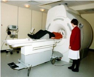
MRI scans are very useful when it comes to detecting the problem with articular cartilage. These machines are very expensive and in most countries there will generally be a huge line just for a couple of scans. This can turn out to be quite expensive for most patients and they might not get their scans done before the damage gets worse for this reason.
Arthroscopy
In this procedure, the surgeon makes a small incision and takes a look inside with a small camera. This camera provides images of the cartilage and these images are graded according to how bad they look starting from 0 to 4.
- 0 – No problem with the cartilage
- 1 – There is some softening in the cartilage and blisters are visible
- 2 – There are small tears on the surface
- 3 – Defect is much deeper
- 4 – Cartilage is almost completely defective and the subchondral bone is exposed
Each grade can be treated in a different fashion and the doctor will explain to the patient the best possible option for him. Once you frequently experience joint pain, you can opt for a foot pain diagnosis and opt for further ankle pain treatment based on the doctor’s advice. Pain in the ankle joint is treated using an ankle fusion surgery.